Stroke and Brain Damage: Location of a Stroke and Symptoms Experienced
A sudden stroke and brain damage due to interrupted blood flow or bleeding in the brain, is a critical neurological event. This damage can occur in various structures of the brain. The symptoms experienced after a stroke are linked to the specific structure damaged and the functions it controls. Various brain structures, their functions, and stroke effects are discussed below.

Stroke in the Left Hemisphere of the Brain
When looking at the brain overall, it is comprised of the left and right hemisphere. There are some generalizations that can typically be made about a stroke in the left brain vs. the right brain. The left hemisphere of the brain is usually dominant for language, math, and logic. Strokes on the left side of the brain may result in:
- Language difficulties: Aphasia, or the inability to understand or produce language, is common. This can include problems with speaking, writing, or reading.
- Right-sided weakness or paralysis: The left side of the brain controls the right side of the body.
- Slow, cautious behavior: Individuals may become more methodical and hesitant.
- Difficulty with numbers and math: Problems with calculations and problem-solving may arise.
- Memory loss: Issues with short-term memory can occur.
Stroke in the Right Hemisphere of the Brain
The right hemisphere is associated with spatial awareness, creativity, and emotional processing. Symptoms of a stroke and brain damage to the right hemisphere may include:
- Left-sided weakness or paralysis: Similar to left brain strokes, but on the opposite side of the body.
- Difficulty with spatial perception: Issues with depth perception, navigation, and understanding visual cues.
- Neglect of the left side: People may ignore the left side of their body or the left side of their environment.
- Impulsive behavior: Decisions may be made hastily without considering consequences.
- Difficulty understanding emotions: Challenges in recognizing and responding to emotions in oneself and others.
It's important to note that these are general characteristics, and individual experiences can vary widely. If the specific structures of the brain that have been affected by a stroke are known, you can begin to glean more information about impairments.
Brain Structures
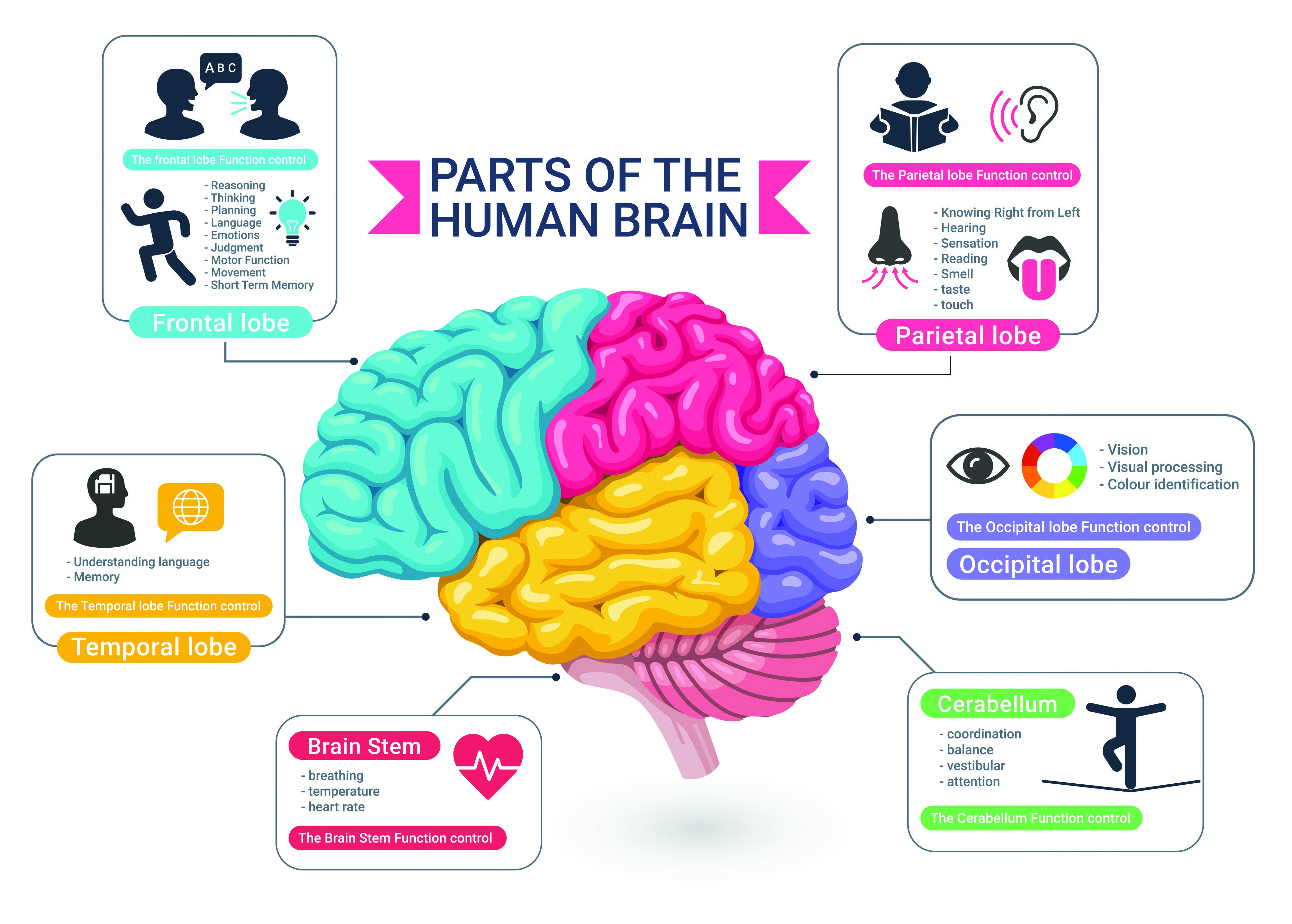
Besides the left and right hemisphere, the brain is further divided into structures called the cerebrum, cerebellum, and the brain stem. The cerebrum contains the frontal, parietal, temporal, and occipital lobes. The brainstem is composed of the midbrain, pons, and medulla oblongata. These structures control different functions of the body. The functions of various parts of the brain are summarized in the infographic below.
Lobes of the Cerebrum
Stroke in the Frontal Lobe
A stroke affecting the frontal lobe can cause problems with motor function, decision-making, problem-solving, and social behavior. Individuals might experience weakness or paralysis on the opposite side of the body, posing challenges with mobility and coordination.
Stroke in the Parietal Lobe
The parietal lobe, when affected, leads to difficulties in recognizing objects or understanding spatial relationships, affecting one's ability to read, write, or do math.
Stroke in the Occipital Lobe
If the stroke occurs toward the back of the brain where the occipital lobes reside, it can lead to visual disturbances, such as loss of half of the visual field or even total blindness.
Stroke in the Temporal Lobe
Should the stroke affect the temporal lobe, it could result in problems with memory, speech, and hearing.
Cerebellum
The cerebellum regulates balance and movement. A stroke in this area can cause loss of balance, difficulty with fine motor skills, unsteady or ataxic gait, and lack of coordination. Cerebellar strokes also may cause vertigo, nausea and vomiting, headache, nystagmus, difficulties swallowing, and slurred speech. Headaches are also not uncommon after this type of stroke.
Brain Stem
The brain stem is composed of the midbrain, pons and medulla oblongata:
- Midbrain: involved in motor control, particularly eye movements, and processing of vision and hearing.
- Pons: helps coordinate face and eye movements, facial sensations, hearing, and balance.
- Medulla Oblongata: regulates vital functions such as breathing, heartbeat, blood pressure, and swallowing.
Brain stem strokes can be devastating having profound effects as this region controls most of the body's automatic functions such as heart rate, breathing, and consciousness.
Signs and symptoms can differ but may include hemiplegia (paralysis on one side of the body) or quadriplegia (paralysis in all four limbs), sensory loss, double vision, dysconjugate gaze (a failure of the eyes to turn together in the same direction), slurred speech, impaired swallowing, decreased level of consciousness, and abnormal respirations. Patients with brain stem strokes may require emergency intubation and mechanical ventilation. Severe consequences can include locked-in syndrome (complete paralysis of all voluntary muscles except for certain eye movements) or brain death.
Blood Supply to the Brain
When discussing where a stroke is located, your MD may indicate that a certain artery was involved. The illustration below shows how vertebral, basilar, and cerebral arteries come together at the base of the brain though there can be anatomical variations among individuals.

Vertebral Arteries
The vertebral arteries run through the neck (cervical vertebral column) and join together at the base of the brain to form the basilar artery which supplies the posterior portion of the brain including the cerebellum and brain stem. The vertebral arteries are sometimes susceptible to dissection
Vertebral artery dissection (VAD) is a relatively rare but significant cause of stroke, particularly in younger and middle-aged patients. A dissection occurs when a tear forms in the inner lining of the artery, allowing blood to enter the arterial wall and cause the wall to balloon out and potentially clot.
This dissection can reduce blood flow to the brain and send clots to smaller vessels, both leading to a stroke. VAD can result from major trauma such as a car accident or even from minor trauma like rapid neck movement or from a sports-related injury. Some causes of VAD have resulted from rollercoaster or amusement park rides, a chiropractic adjustment of the neck, and even getting ones hair washed at the beauty parlor (see beauty parlor syndrome). In other cases, it may occur spontaneously without an identifiable cause.
Symptoms of VAD can vary, but often include neck pain and headache, dizziness, loss of balance, and potentially more serious symptoms like difficulty speaking or swallowing.
Basilar Artery
Inside the skull, the two vertebral arteries join up to form the basilar artery. The basilar artery is the main blood vessel that forms the posterior circulation of the brain. A stroke in the basilar artery can cause speech difficulties, visual disturbances, cranial nerve palsies, nausea and vomiting, vertigo and altered consciouscness.
Cerebral Arteries
Stroke in the Middle Cerebral Artery (MCA)
The middle cerebral artery (MCA) is the most common cerebral occlusion site. The MCA is a complex artery with several branches, each supplying blood to specific areas of the brain. A stroke affecting one of these branches can lead to a variety of symptoms depending on the location of the blockage.
MCA Branches:
M1 segment: The proximal portion of the MCA before it divides into smaller branches. Strokes in this area may cause extensive brain damage due to the large amount of brain tissue supplied by the M1. Occlusion often leads to severe neurological deficits.
- Common symptoms: Severe weakness or paralysis on the opposite side of the body, severe aphasia (difficulty with speech or understanding language), visual disturbances, and sensory loss.
M2 segment: The middle portion of the MCA where it begins to branch. Because the M2 segment supplies smaller areas of the brain, symptoms tend to be more specific.
- Possible symptoms: Depending on the specific branch affected, symptoms can include facial weakness, arm weakness, hand weakness, sensory loss, aphasia, or neglect of one side of the body.
M3 & M4 segment: The distal branches of the MCA supplying specific regions of the brain. As the smallest branches of the MCA, occlusions here typically result in very specific deficits.
- Possible symptoms: Can include isolated weakness or numbness in a specific body part, mild aphasia, or visual disturbances.
It's important to note that these are general descriptions and individual experiences can vary widely. The severity of symptoms and the specific areas affected by the stroke will influence the overall impact on a person's life.
Stroke in the Posterior Cerebral Artery (PCA)
The PCA supplies blood to the occipital lobe which is responsible for vision, as well as parts of the thealamus, which is involved in sensory processing. A stroke and brain damage in the posterior cerebral artery (PCA) can cause a variety of symptoms, primarily affecting vision and sensory perception.
Common effects of a PCA stroke include visual disturbances such as:
- Homonymous hemianopia: Loss of vision in one half of the visual field in both eyes.
- Cortical blindness: Complete loss of vision.
- Visual hallucinations: Seeing things that aren't there.
- Difficulty recognizing faces (prosopagnosia).
- Difficulty perceiving colors.
Sensory symptoms may occur as well and can include:
- Numbness or tingling in the body.
- Pain (thalamic pain syndrome).
Other potential symptoms:
- Dizziness or vertigo.
- Difficulty with balance.
- Memory problems.
- Language difficulties.
- Motor weakness (though less common than in strokes affecting other arteries).
Stroke in the Anterior Cerebral Artery
The anterior cerebral artery (ACA) supplies the anterior medial portions of the frontal and parietal lobes. Classic signs of an ACA stroke are contralateral leg weakness and sensory loss.
Other Brain Structures Stroke and Brain Damage
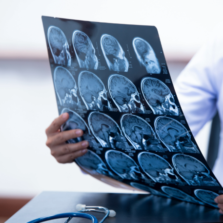
Stroke and brain damage can occur in other brain structures including the thalamus, basal ganglia, internal capsule, hypothalamus, amygdala, and hippocampus.
- Thalamus - A stroke in the thalamus may result in sensory changes, debilitating pain (central post-stroke pain syndrome), balance issues as well as symptoms common to strokes in other areas of the brain.
- Basal Ganglia - The basal ganglia is associated with motor control, learning, habit formation, and cognition. A stroke in the basal ganglia can result in motor control problems as well as changes in cognition and behavior.
- Internal Capsule - The internal capsule helps control movement. A stroke and brain damage in this area will often result in motor control problems including hemiplegia (paralysis on one side of the body), hemiparesis (weakness on one side of the body), and facial weakness.
- Hypothalamus - The hypothalamus regulates bodily functions like temperature, sleep, and hunger. A hypothalamic stroke may result in hormone dysfunction, difficulty regulating body temperature, sleep disturbances, and appetite changes.
- Amygdala - The amygdala is involved in emotional processing, especially for fear and aggression. A stroke in the amygdala may result in significant behavioral changes including difficulty controlling emotions, excessive anxiety, unfound phobias, anger, depression, and social difficulties. Memory changes may occur as well .
- Hippocampus - A stroke in the hippocampus can result in difficulty forming new memories, difficulty recalling past events, and impaired spatial navigation. One may struggling to recognize familiar places.
These are some of the brain structures that may be affected by stroke but is not an exhaustive list. Stroke patients should consult with their doctor regarding what specific area of their brain was affected and what symptoms might be expected.
If you'd like to learn more about brain anatomy, stroke and brain damage; visit https://mayfieldclinic.com/pe-anatbrain.htm#
Get Our Stroke Rehab Guide
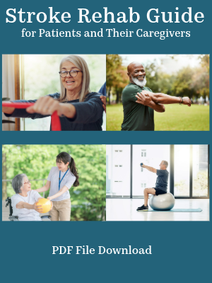
Our comprehensive stroke rehab guide in pdf format is designed for both patients and caregivers who want clear, practical ways to support recovery, improve daily function, and regain independence at home. It includes
- Rehab exercises with pictures for safe home practice
- Physical, occupational, and speech therapy guidance
- Tips for daily activities and adaptive equipment
- Answers to common questions from patient and caregivers
- Information on stroke causes, treatment, and prevention
A single therapy visit can run $150 or more. The Stroke Rehab Guide is only $14.99, and includes a pdf guide you can continue to refer to in the future with exercises and information on stroke recovery. In addition, any time an update or new version of the guide is written, you will get the updated version for free.
About the Author
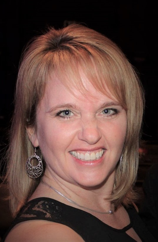
Karen Murray, OTR, CHT, CSRS, is a licensed occupational therapist, Certified Stroke Rehabilitation Specialist, Certified Hand Therapist, and Certified Personal Trainer with over 29 years of experience working with stroke survivors in hospital, outpatient, and home settings. She founded Stroke-Rehab.com to help patients and caregivers better understand stroke recovery, find evidence-based resources, and regain independence at home.
Medical Disclaimer: All information on this website is for informational purposes only. This website does not provide medical advice or treatment. Always seek the advice of your physician or other healthcare provider before undertaking a new healthcare or exercise regimen. Never disregard professional medical advice or delay seeking medical treatment because of something you have read on this website. See the disclaimer page for full information.
